
×
![Enlarged Image]()
Integrated Computational Materials Engineering (ICME)
Automated Region Growing Technique for EFTEM images - Example
Author(s): Mark A. Tschopp, G.B. Viswanathan, J.S. Tiley
Corresponding Author: Mark Tschopp
Here is a quick tutorial on how to use this code:
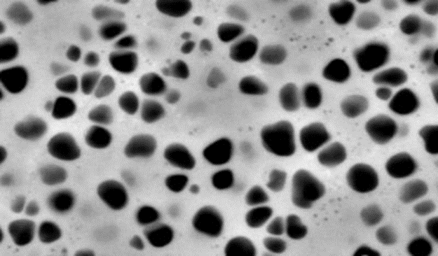
1. Open up segment_master.m in MATLAB. 2. Run the first portion of the MATLAB script. MATLAB will pop-up a box to select the image in a tif format. Select the image file. I have selected the following image for this example.
Abstract
Region growing technique for segmenting microstructural features in an automated way, as first used for energy-filtered transmission electron microscopy images[1][2] The use of different electron loss edges in energy filtered transmission electron microscopy (EFTEM) has allowed researchers to capture images of the morphology and size of precipitates in nickel-based superalloys. In this work, we discuss a computational methodology for automated detection of secondary and tertiary γ’ precipitates in EFTEM images. The optimum parameters for the automated region growing technique were identified using a combination of visual inspection and intensity information from the EFTEM images. The microstructural statistics obtained from the segmented γ’ precipitates agreed with those of the manually segmented precipitates. Then, automated segmented precipitates are used to extract microstructural information about the distributions of equivalent diameters of 656 tertiary precipitates along with the distances to the nearest secondary precipitates. The significance of this technique is its ability to automate segmentation of precipitates in a reproducible manner for acquiring microstructural statistics that relate to both processing and properties.Description
This code is based on the region growing algorithm available at MATLAB Central.Here is a quick tutorial on how to use this code:
1. Open up segment_master.m in MATLAB. 2. Run the first portion of the MATLAB script. MATLAB will pop-up a box to select the image in a tif format. Select the image file. I have selected the following image for this example.
3. The image will open up in a new window. At this point, you can crop the image. Drag a box around the region that you would like to analyze (or select a box around the entire image) and double click inside the image. The image now looks like this and MATLAB has saved this cropped image as a new file.
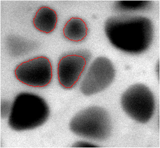
4. Now comes the region growing portion of the script. Run this portion as many times as needed. This portion will open up the cropped image and allow the user to select a box around the microstructural feature of interest. Bound the particle of interest and then hit enter. Now, select a seed point within the particle and hit enter. The region growing algorithm will now execute and will output the segmented image to a binary matrix (1's are particles, 0's are matrix) and the time the algorithm took to the screen (the bigger the box and the particle, the longer the time it takes).

5. When all the particles are segmented, run the third portion of the code. This will save the binary image of the segmented particles and an image that shows the boundaries of the segmented particles on the original intensity image.
6. All done. There are a number of modifications that you can make to this routine to suit the particular application.
Conclusions
In this work[1][2]we discuss a computational methodology for automated detection of secondary and tertiary γ’ precipitates in energy filtered transmission electron microscopy (EFTEM) images. Several important parameters for the automated region growing technique were investigated using a combination of visual inspection and intensity information from the EFTEM images. These parameters were related to the method used for calculating the stopping criterion, the local density weighting function, and the seed point selection method. After optimizing these parameters, the microstructural statistics obtained from the γ’ precipitates segmented with the automated technique were compared with the same precipitates segmented manually. On average, the results show that the precipitate area (equivalent diameter) obtained using the automated technique is approximately 14% (7%) lower than that of the same precipitates segmented manually. The automated region growing technique presented here is suitable for detecting secondary and tertiary γ’ precipitates of complex morphology and varying intensity contrast in a reproducible manner.This technique was then used to segment the secondary and tertiary γ’ precipitates from EFTEM images of a single crystal nickel-based superalloy that was slow cooled after aging for 200 hours. The segmented precipitates were used to calculate average/extreme microstructural statistics for this processing condition. In addition to calculating the distribution of tertiary precipitate equivalent diameters, the distribution of distances from 656 tertiary precipitates to the nearest secondary precipitates was calculated. The extreme values of the distribution shows that the majority of tertiary γ’ precipitates are located greater than 50 nm and less than 300 nm from the secondary γ’ precipitate surfaces for this processing condition. The significance of this technique is its ability to automate segmentation of precipitates in a reproducible manner for acquiring microstructure statistics that relate to both processing and properties.
Acknowledgments
MAT would like to acknowledge funding through the visiting scientist contract at the Air Force Research Laboratory on "Damage Mechanics Model Development for Monocrystalline Superalloys."References
- ↑ Tschopp, M.A., Tiley, J.S., Viswanathan, G.B., "Automated Identification and Characterization of Secondary & Tertiary Gamma Prime Precipitates in Nickel-based Superalloys," Materials Science & Technology 26(12) (2010) 1414-1422, https://www.tandfonline.com/doi/abs/10.1179/026708309X12560332736638
- ↑Tiley, J.S., Rajagopolan, S., Viswanathan, G.B., Shiveley, A., Tschopp, M.A., Banerjee, R., Fraser, H.L., "Measurement of Gamma Prime Precipitates in a Nickel-based Superalloy using Energy-Filtered Transmission Electron Microscopy coupled with Automated Segmenting Techniques," Micron, 41(6) (2010) 641-647, https://www.sciencedirect.com/science/article/pii/S0968432810000545?via%3Dihub