
Integrated Computational Materials Engineering (ICME)
Turtle shell
Abstract
The multiscale structure, materials properties, and mechanical responses of
the turtle shell (Terrapene carolina) were studied to understand the
fundamental knowledge of naturally occurring biological penetrator-armor
systems. The structure observation and chemical analysis results revealed that
the turtle shell carapace comprises a multiphase sandwich composite structure
of functionally graded material having exterior bone layers and a foam-like
bony network of closed-cells between the two exterior bone layers. Although the
morphology was quite different, the exterior bone layers and interior bony
network possessed comparable hardness and elastic modulus values of ~1 GPa and
~20 GPa, respectively. Compression and flexure test results showed a typical
nonlinear deformation behavior recognizant of man-made foams. The mechanical
test results revealed that the interior closed-cell foam layer plays a
significant role on the overall deformation behavior of the turtle shell. The
finite element analysis simulation results showed comparable agreement with the
actual experimental test data. This systematic study could provide fundamental
understanding for structure-property phenomena and biological pathways to
design bio-inspired synthetic composite materials
Author(s): Hongjoo Rhee, Mark F. Horstemeyer, Y. Hwang, H. Lim, H. El Kadiri,
W. Trim
Corresponding Author: Hongjoo Rhee
Structure
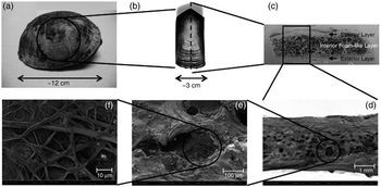
Fig. 1. Multiscale hierarchy and structure of the turtle shell; (a) a morphology of the turtle shell carapace, (b) a costal scute showing the successive growth pattern, (c) a crosssectional view of the carapace showing composite layers, (d) an SEM micrograph of a fracture surface, (e) an SEM micrograph of a cell structure, and (f) an SEM micrograph of a fibrous structure inside of the cell.
Structure observations on the turtle shell revealed a multiphase composite
material that is arranged by a multiscale hierarchy. Such a multiscale
hierarchical structure of the turtle shell carapace is depicted in Fig. 1. The
turtle shell comprises a series of connected individual plates covered with a
layer of horny keratinized scutes (Fig. 1a–b). The scutes are made up of a
fibrous protein called keratin that also comprises the scales of other reptiles
[1]. These scutes overlap the seams
between the shell bones and serve to reinforce the overall protection to the
shell. The carapace is made of a sandwich composite structure of functionally
graded material (FGM) having relatively denser exterior layers and an interior
fibrous foam-like layer (Fig. 1c–d). SEM micrographs clearly revealed such
fibrous structure inside of the cell (Fig. 1e–f).
The internal structure of the turtle shell was nondestructively observed by
using an X-ray computed tomography (CT) and obtained images are provided in
Fig. 2. The X-ray CT was carried out by using a v|tome|x by phoenix|x-ray. The
X-ray CT images clearly showed that the pores within the interior foam-like
layer of the turtle shell carapace were closed-cell type and randomly
distributed. In addition, the results obtained from the in-house image analyzer
software revealed that the porosity levels of the relatively denser exterior,
interior foamlike layer, and whole turtle shell carapace including all three
layers were 6.86%, 65.5%, and 48.9%, respectively.
Figs. 3 and 4 show the microstructure observation and chemical analysis results
obtained from various surfaces of the turtle shell. Three different layers of
the outermost keratin layer, right underneath the keratin layer, and the inside
surface of the turtle shell carapace were observed and analyzed by using an SEM
and an energy dispersive X-ray (EDX) spectroscopy technique, respectively.
These layers have different surface microstructures and chemical compositions.
The EDX analysis showed that the outermost keratin layer mainly consists of
carbon (C), oxygen (O), nitrogen (N), and sulfur (S) that are main constituents
of the protein. The result is not surprising since the keratins are a family of
fibrous structural proteins, also called scleroproteins. Unlike the outermost
keratin layer, right underneath the keratin layer and the inside surface of the
turtle shell carapace contained abundant additional minerals as indicated by
the presence of calcium (Ca, 15–20 wt.%), phosphorous (P, 7–10 wt.%), sodium
(Na), chlorine (Cl), and magnesium (Mg) that are known to be main components of
the bone.
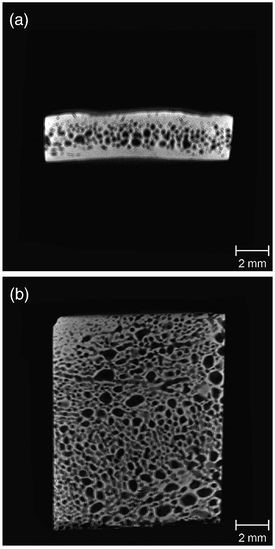
Fig. 2. (a) A side sectional view and (b) a top sectional view of the turtle shell carapace coupon obtained from X-ray CT single slice scan showing randomly distributed closed-cell pores within the foam-like interior layer.
Fig. 3. SEM micrographs obtained from different surfaces of the turtle shell carapace; (a) the outermost keratin layer, (b) underneath the keratin layer, and (c) inside surface.

Fig. 4. Chemical analysis results obtained from different surfaces of the turtle shell carapace; (a) the outermost keratin layer, (b) underneath the keratin layer, and (c) inside surface.
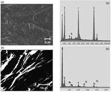
Fig. 5. SEM micrographs obtained from the fracture surface of the turtle shell carapace; (a) bony layers and (b) inside of the closed-cell (fibers). Fig. 6. Chemical analysis results obtained from the fracture surface of the turtle shell carapace; (a) bony layers and (b) inside of the closed-cell (fibers).
The microstructures and chemical analysis results obtained from different locations of the fracture surfaces of the turtle shell carapace are provided in Figs. 5 and 6. The chemical compositions obtained from the exterior layers and the network (e.g. closed-cell wall) region within the foam-like interior layer were quite similar to those can be found in Fig. 4b–c. The fibers inside of the closedcell also showed an accordant chemical composition (Fig. 6b), which implies that they include “bony” fibers. The microstructure observation and chemical analysis results obtained from various locations of the turtle shell clearly revealed that the turtle shell carapace is made of a sandwich composite structure having exterior lamellar bone layers and an interior bony network of closedcell fibrous foam layer.
Mechanical Properties
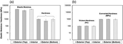
Fig. 7. Indentation test results obtained from (a) nano-indentation and (b) Vickers hardness tests on the side surface of the turtle shell carapace.
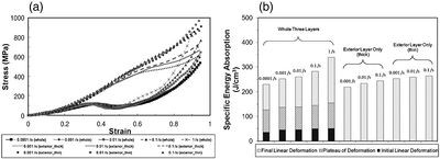
Fig. 8. Quasi-static compression test results on the turtle shell carapace coupon specimens under various strain rates and specimen geometries; (a) stress versus strain curves and (b) specific energy absorption as a function of density.
Experimental results obtained from the nano- and microindentation tests on
the side surfaces of the turtle shell carapace are provided in Fig. 7. The
results showed that the exterior layers and interior bony closed-cell walls
possess comparable hardness and modulus values. Hardness and elastic modulus
values obtained from the nano-indentation tests ranged from 0.8–1.1 GPa and
18.3– 24.8 GPa, respectively; whereas, the average hardness value obtained from
the Vickers hardness tests was about Hv100 that corresponds to 0.98 GPa. There
were small variations in hardness and elastic modulus values from experiments
due to the roughness of the specimen. The nano-indentation test results reflect
highly localized micromechanical properties that may contain porous or
impurities in its texture. Since the regions of indentation are so small that
local impurities or defects can induce uncertainties in the measurements. This
effect is minimized under Vickers hardness test set-up and the exterior layers
and closed-cell walls within an interior layer possessed comparable hardness
values.
For quasi-static compression tests, two different types of coupon specimens
including all three layers and then only a bony exterior layer were prepared.
The effect of strain rate on the mechanical behavior of the turtle shell was
compared with respect to the different density levels and the raw data obtained
from the tests is illustrated in Fig. 8a. The lower five curves (represented by
lines with symbols) were obtained from the test specimens including all three
layers (two exterior and an interior layers); whereas the upper six curves were
obtained from the specimens only containing a relatively denser exterior layer.
Top three curves (in symbols) among those six curves were obtained from thinner
specimens and the bottom three curves (in lines) represent thicker specimens.
The thickness difference between those two regimes was about 15%. The favorable
deformation mechanism of the turtle shell carapace under quasi-static
compression test conditions can be explained by importing that of synthetic
foams and/or honeycombs since fundamental structures of the test specimens are
similar to those of such cellular solids. At small strains, the specimens were
deformed in a linear elastic manner due to the cell wall bending[2]. Soon after
the initial linear elastic deformation, a plateau of deformation was reached,
because of the buckling of the cell walls. After such a plateau of deformation,
another period of linear deformation was proceeded since a densification
occurred resulting in a rapid increase of compressive stress. When comparing
the specimens containing the exterior region only, the thicker specimens showed
a similar deformation yet much weaker behavior than those can be observed in
the specimens including all three layers; whereas the thinner specimens showed
almost a linear compressive deformation behavior simply because of the density
and structure differences. Most of discernible pores within exterior layer are
distributed near the region between the exterior layer and interior foam-like
layer. Fig. 8b provides the comparison of specific energy absorption obtained
from the quasi-static compression test results (Fig. 8a). Density and porosity
levels of the test specimens were already considered in this normalized data.
The energy absorption ability of the turtle shell carapace increased with
increasing strain rate for a given density level. The composite layers
including all three layers showed better energy absorption ability compared to
the exterior layer for any given strain rate. In addition, such composite
layers possessed a considerable amount of plateau of deformation that is a
model index of good energy absorbing materials. The combining information of
these two plots in Fig. 8 is very important to design the optimum energy
absorbing composite material. For example, composite foam materials can be
tailored to give the best combination of properties for a given package by
choosing the right combination of the cell wall materials, relative density,
reinforcement phases, and so on.
Reference
1. D.R Katti, S.M. Pradhan, K.S. Katti, Rev. Adv. Mater Sci. 6(2004)
162.
2. L.J. Gibson, M.F. Ashby, Cellular Solids: Structure and Properties – Second
Edition. Cambridge University Press, Cambridge, U.K., 1997.
3. Citation: H. Rhee, M.F. Horstemeyer, Y. Hwang, H. Lim, H. El Kadiri, and W.
Trim, “A study on the structure and mechanical behavior of the Terrapene
carolina carapace: a pathway to design bio-inspired synthetic composites,”
Materials Science and Engineering C 29 (2009) 2333-2339.