
Integrated Computational Materials Engineering (ICME)
ICME Overview of Mechanical Properties of the Lipid Bilayer during Traumatic Brain Injury
Background
Traumatic brain injury (TBI) has become a concern for many nations around the world. Defined as an alteration in brain function or pathology due to some external force or acceleration, TBI includes an expansive number of injury types and can be manifested as many symptoms including death [1]. Because of the wide ranging symptoms, every year both human and economic losses result from the millions of individuals who suffer from TBI [2]. Despite these losses, much is still yet to be understood due to the complexity of TBI, difficulty in diagnosing and studying mechanisms in vivo, and the time scales involved in different injury forms. Therefore, understanding the phenomenon of TBI across multiple scales is essential to expanding the current physical knowledge of the brain and brain injury. This understanding will allow for higher fidelity models of the human brain and result in better diagnostic tools for the medical field. The five length scales and necessary bridges can be seen in Figure 1.
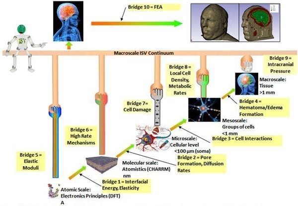
Figure 1. Multiscale diagram displaying the necessary scales and information passed across the bridge scales.
Macroscale
Due to damage on lower scales during TBI, the brain often develops an increased intracranial pressure resulting from hematoma and edema, two forms of fluid buildup, formation. This can lead to an increased life risk due to more pressure being placed on the brain and neurons and can cause further cell death [3]. In order to determine how much risk of edema and hematoma formation, the cell interactions must be incorporated from the mesoscale.
Mesoscale
The mesoscale needs to incorporate if cell death occurs on the microscale
because dying cells can release excitotoxic molecules into the extracellular
space. These molecules then cause cellular damage to other cells by
overstimulating receptors that allow an influx of calcium ions into the
neighboring cells. The calcium ion influx then can activate several proteases
that will damage the cells [4].
This will cause the death of only a few cells initially to spread and create a
region of dead cells. This damage can cause swelling resulting in hematomas and
edema [3]. The amount of cell death can be measured using methods such as local
cell density and metabolic rates because higher cell death will result in lower
local cell densities and metabolic rates [5].
This is due to the fact the damaged cells will proceed through apoptosis,
neurosis, or a combination of the two and the cells will discontinue
metabolizing after death occurs.
Microscale
The microscale requires information about if the cellular structures are
damaged on the molecular scale. In this case, only the lipid bilayer portion of
the cell membrane is being examined. If damage to the cell membrane does occur,
excitotoxic calcium ions can enter the cell resulting in increased
intracellular ion concentrations. These will activate cysteine protease
calpains, calcium-activated neutral proteases, which cleave the cytoskeletal
protein spectrin resulting in a failure of the cellular cytoskeleton [4].
This will cause the release of more excitotoxic ions and molecules into the
extracellular space. These will then affect neighboring cells as mentioned in
the mesoscale.
Molecular Scale
The molecular scale requires information from the atomistic scale as to how
the molecules will interact with one another. This allows for a simulation of
many of the molecules to be run giving proper behavior. The molecular scale
will pass up the high rate mechanisms related to the lipid bilayer. This will
determine if the bilayer is damaged during the scenarios, such as blast waves,
being inspected. To determine if the bilayer is damaged, molecular dynamics are
used to determine if pore formation occurs and if the excitotoxic ions of
interest can enter the cell. Additionally, diffusion rates at which the ions
enter the cell can also be determined using molecular dynamics [6].
These damage criteria will be passed up to the microscale to determine if cell
death occurs.
Electronic Principle Scale
In order to perform the molecular dynamics needed for the lipid bilayer, the interfacial energy between the two layers of lipids and the interfacial energy of the lipid water interface must be determined using density functional theory. Additionally, the elasticity between the molecules must be determined for bonding purposes.
Reference
1. Menon, D. K., Schwab, K., Wright, D. W., & Maas, A. I. (2010). Position
Statement: Definition of Traumatic Brain Injury. Archives of physical medicine
and rehabilitation, 91(11), 1637-1640.
2. Sabet, A., Christoforou, E., Zatlin, B., Genin, G. M., & Bayly, P. V.
(2008). Deformation of the human brain induced by mild angular head
acceleration. [Research Support, N.I.H., Extramural Research Support, Non-U.S.
Gov't]. J Biomech, 41(2), 307-315. doi: 10.1016/j.jbiomech.2007.09.016
3. 3.0 3.1 Prins, M. L., Lee, S. M., Cheng, C. L. Y., Becker, D. P., & Hovda,
D. A. (1996). Fluid percussion brain injury in the developing and adult rat: a
comparative study of mortality, morphology, intracranial pressure and mean
arterial blood pressure. Developmental Brain Research, 95(2), 272-282. doi: 10.1016/0165-3806(96)00098-3
4. 4.0 4.1 Farkas, O., & Povlishock, J. T. (2007). Cellular and subcellular
change evoked by diffuse traumatic brain injury: a complex web of change
extending far beyond focal damage. In T. W. John & I. R. M. Andrew (Eds.),
Progress in Brain Research (Vol. Volume 161, pp. 43-59): Elsevier.
5. Giza, C. C., & Hovda, D. A. (2001). The Neurometabolic Cascade of
Concussion. J Athl Train, 36(3), 228-235.
6. Wang, J., & Hou, T. (2011). Application of molecular dynamics simulations in
molecular property prediction II: diffusion coefficient. J Comput Chem, 32(16),
3505-3519. doi: 10.1002/jcc.21939.
Epub 2011 Sep 22.